Volume 27, Number 2—February 2021
Research Letter
Protective Immunity and Persistent Lung Sequelae in Domestic Cats after SARS-CoV-2 Infection
Abstract
Severe acute respiratory syndrome coronavirus 2 readily transmits between domestic cats. We found that domestic cats that recover from an initial infection might be protected from reinfection. However, we found long-term persistence of inflammation and other lung lesions after infection, despite a lack of clinical symptoms and limited viral replication in the lungs.
Previous studies have demonstrated the transmissibility of severe acute respiratory syndrome coronavirus-2 (SARS-CoV-2) by direct or indirect contact between domestic cats (1,2). Given the close relationship between cats and humans, further characterization of the biology of SARS-CoV-2 in cats is warranted.
We inoculated domestic cats with SARS-CoV-2, and on postinfection days 3, 6, and 10, sampled organs to titrate virus (Appendix Figure 1). In plaque-forming assays in VeroE6/TMPRSS2 cells, infectious viruses were detected in the nasal turbinates and trachea of all animals on day 3, and most on day 6, whereas virus detection in the lungs was limited on day 3 and absent on day 6 (Appendix Figure 2, panel A). These results suggest that the virus replicated efficiently in upper respiratory organs, which might contribute to its high transmissibility among cats. Infectious virus was cleared from the upper and lower respiratory organs by day 10 (Appendix Figure 2, panel A). No animal showed any signs of respiratory illness during the study (Appendix Figure 3). Infectious virus was not detected (detection limit 10 pfu/g of tissue) in other examined organs (e.g., brain, liver, spleen, kidney, small and large intestine, heart, and eyelids). Viral antigen was detected in nasal turbinates and trachea but was sparse within the lungs at day 3 (Appendix Figure 4).
We conducted histopathologic examination of the lungs, trachea, and nasal turbinates. Lymphocytic inflammation within the tracheal submucosa was present on days 3 to 10, whereas lymphocytic to mixed inflammation in the nasal cavity was more severe on days 3 and 6 but minimal on day 10. In lungs, moderate lesions persisted despite clearance of virus. On day 3, we observed mild bronchitis with lymphoid hyperplasia, moderate to severe histiocytic bronchiolitis with partial to complete occlusion of lumina, and moderate to severe thickening of alveolar septa (Appendix Figure 2, panel B; Appendix Figures 4, 5). Interstitial inflammatory infiltrate decreased significantly over time (p = 0.0012, F = 34.70, by 1-way analysis of variance) (Appendix Figure 2, panel C); however, by day 10, alveolar septa remained thickened (Appendix Figure 5). Bronchiolitis remained with partial occlusion of bronchioles, even in regions with minimal alveolar lesions (Appendix Figure 2, panel B).
Because SARS-CoV-2 did not cause acute lethal respiratory disease in the cats in our study, cats are a compelling animal model for studying the long-term effects of nonfatal infections. Cats were infected with SARS-CoV-2 and euthanized at postinfection day 28 (Appendix Figure 6, 7). Persistent lung lesions were observed 28 days after infection, including histiocytic bronchiolitis with luminal plugs and thickened alveolar septa, similar to lesions observed on day 10 but with more chronic features such as peribronchiolar fibrosis and vascular proliferation within the thickened interstitium. We observed a notable dearth of fibrosis within alveolar septa, in contrast to what has been reported for humans with severe acute respiratory syndrome or Middle East respiratory syndrome (3,4). One cat had severe pneumonia with fibrin in alveolar spaces and endothelialitis (Appendix Figure 8), similar to what has been reported in humans with fatal coronavirus disease (5), although this cat did not show any respiratory signs.
Figure 1
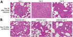
Figure 1. Comparison of histopathology between cats on day 28 after initial infection with severe acute respiratory syndrome coronavirus 2 and on day 21 after reinfection. Bronchioles and alveoli of cats (cats...
Figure 2
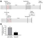
To determine whether previous infection provides protection from future potential infection by SARS-CoV-2, we performed a reinfection study with 2 groups of cats. We previously reported that SARS-CoV-2 was transmitted from cats inoculated with the virus to cohoused, naive cats (1). In the previous study, the 3 cats that had been inoculated with SARS-CoV-2, whose nasal swabs were virus-negative on day 6 or 7 after the initial infection (1), were reinoculated with the same virus 4 weeks after the initial infection (Figure 1; Figure 2, panel A). No infectious virus was detected in the nasal or rectal swabs after reinfection, suggesting that the animals were protected from reinfection. These cats were euthanized at 21 days after reinfection (49 days after the initial infection), and tissue was submitted for histopathologic examination. The reinfection group showed lesions that were comparable with lung lesions observed on day 28 but with less severe thickening of alveolar septa (p = 0.041, by unpaired t-test) (Figure 1; Figure 2 panel B). The 3 cats in the other group, which recovered from infection that was transmitted by contact with virus-inoculated cats, were reinfected with the virus at ≈4 weeks (29–32 days) after transmission. On day 3 after reinfection, organs were harvested; infectious virus was not detected (detection limit 10 pfu/g of tissue) in respiratory organs or other organs analyzed (e.g., brain, liver, spleen, kidney, small and large intestine, heart, and eyelids). These results suggest that virus infection by natural transmission between cats, as well as by experimental inoculation, induces protective immunity against a second SARS-CoV-2 infection.
In conclusion, SARS-CoV-2 replicated effectively in the upper respiratory tract in cats, and infectious virus was cleared from the lungs within 6 days of infection; however, histopathologic examination demonstrated chronic lung sequelae in cats even a month after viral clearance. After initial infection with SARS-CoV-2, cats were protected from reinfection, with no virus replication in respiratory organs and no additional lung damage.
Dr. Chiba is a molecular virologist at the Influenza Research Institute at the University of Wisconsin–Madison, with a background in innate immunity studies and structural biology. Her primary research interests include mechanisms of virus infection, virus antigenicity, and host immune responses.
Acknowledgments
We thank Gillian McLellan for the cats used in this study and Sue Watson for scientific editing. We would also like to thank Angela Brice and Olga Gonzalez for sharing their expertise with our pathologists during consultation as well as Amanda Novak, Emily Tran, and Sara Stuedemann for their technical support.
This research was supported by the Center for Research on Influenza Pathogenesis, funded by the National Institutes of Allergy and Infectious Diseases, National Institutes of Health (grant no. HHSN272201400008C awarded to Y.K); the Research Program on Emerging and Re-emerging Infectious Disease from Japan Agency for Medical Research and Development (AMED) (grant no. JP19fk0108113 awarded to Y.K.); the Japan Initiative for Global Research Network on Infectious Diseases from AMED (grant no. JP19fm0108006 awarded to Y.K.); the Japan Program for Infectious Diseases Research and Infrastructure from AMED (grant no. JP20wm0125002 to Y.K.); and a University of Wisconsin K12 Career Development Award from the National Institute of Diabetes and Digestive and Kidney Diseases (grant no. K12DK100022 awarded to L.K.C.).
References
- Halfmann PJ, Hatta M, Chiba S, Maemura T, Fan S, Takeda M, et al. Transmission of SARS-CoV-2 in domestic cats. N Engl J Med. 2020;383:592–4. DOIPubMedGoogle Scholar
- Shi J, Wen Z, Zhong G, Yang H, Wang C, Huang B, et al. Susceptibility of ferrets, cats, dogs, and other domesticated animals to SARS-coronavirus 2. Science. 2020;368:1016–20. DOIPubMedGoogle Scholar
- Cheung OY, Chan JW, Ng CK, Koo CK. The spectrum of pathological changes in severe acute respiratory syndrome (SARS). Histopathology. 2004;45:119–24. DOIPubMedGoogle Scholar
- Das KM, Lee EY, Singh R, Enani MA, Al Dossari K, Van Gorkom K, et al. Follow-up chest radiographic findings in patients with MERS-CoV after recovery. Indian J Radiol Imaging. 2017;27:342–9. DOIPubMedGoogle Scholar
- Ackermann M, Verleden SE, Kuehnel M, Haverich A, Welte T, Laenger F, et al. Pulmonary vascular endothelialitis, thrombosis, and angiogenesis in Covid-19. N Engl J Med. 2020;383:120–8. DOIPubMedGoogle Scholar
Figures
Cite This ArticleOriginal Publication Date: January 07, 2021
Table of Contents – Volume 27, Number 2—February 2021
| EID Search Options |
|---|
|
|
|
|
|
|
Please use the form below to submit correspondence to the authors or contact them at the following address:
Yoshihiro Kawaoka, 575 Science Dr, Madison, Wisconsin 53711, USA
Top